Introduction
Bladder exstrophy repair techniques have evolved over the years. The most frequently utilized approaches include Complete Primary Reconstruction of Exstrophy (CPRE), Modern Staged Repair of Exstrophy (MRSE), and the Kelly Radical Soft Tissue Mobilization technique.1 A successful repair of bladder exstrophy aims to achieve closure of the bladder plate and the anterior abdominal wall, and to reconstruct functional and cosmetically acceptable genitalia while maintaining a low-pressure, competent bladder to ultimately achieve continence.2 To achieve a tension-free closure, pelvic osteotomy is recommended during the initial repair; however, some institutions have reported good results without osteotomy.1
There is currently no consensus on the routine use of pelvic osteotomy in the repair of bladder exstrophy, primarily because of concerns about the associated morbidity, a lack of osteotomy expertise and operative infrastructure, and inadequate post-operative orthopaedic support including immobilization requirements such as splints, pelvic binders, and external fixators, especially in settings with limited resources.3 In addition, the increased operative time (approximately 45–90 additional minutes) makes it a less desirable option for many centres.4
The use of a rectus abdominis flap has been shown to offer a technically feasible alternative for bladder exstrophy repair, as it can be performed without pelvic osteotomy. This case series aims to document our experience with 8 cases managed using a modified approach incorporating a rectus abdominis flap without osteotomy. Specifically, we highlight the technique employed and the short-term outcomes in an African population with bladder exstrophy.
Patient Selection and Description
Between 2022 and 2025, 8 patients with classic bladder exstrophy were surgically managed at the Kenyatta National Hospital. Seven were male patients, and 1 was female. All patients received a standard clinical evaluation and screening for associated anomalies, including an abdominal and pelvic ultrasound. All patients underwent primary closure of bladder exstrophy using a rectus abdominal flap technique. All surgeries were performed by a single surgeon specialized in the field of paediatric urology. The patient details are summarised in Table 1.
Surgical Procedure
The general principles of this procedure are based on Gearhart’s three-stage MRSE, with the added modification of using a rectus abdominis flap for abdominal wall closure during the first stage.
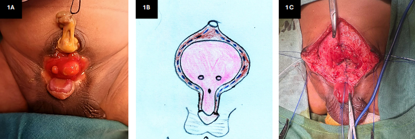
The procedure is performed under general anaesthesia with a caudal block for perioperative pain control. The patient is positioned supine, and site preparation is done from the abdomen to the level of the knees. The umbilicus is routinely maintained for retraction purposes, which is achieved by applying a 3/0 monofilament stay suture as shown in Figure 1A.
Incisions are made along the margins of the bladder using both a surgical blade and electrocautery. Cranially, these incisions extend to the level of the umbilicus, while caudally they extend on either side of the urethral plate to a point distal to the verumontanum, as illustrated in Figure 1B.
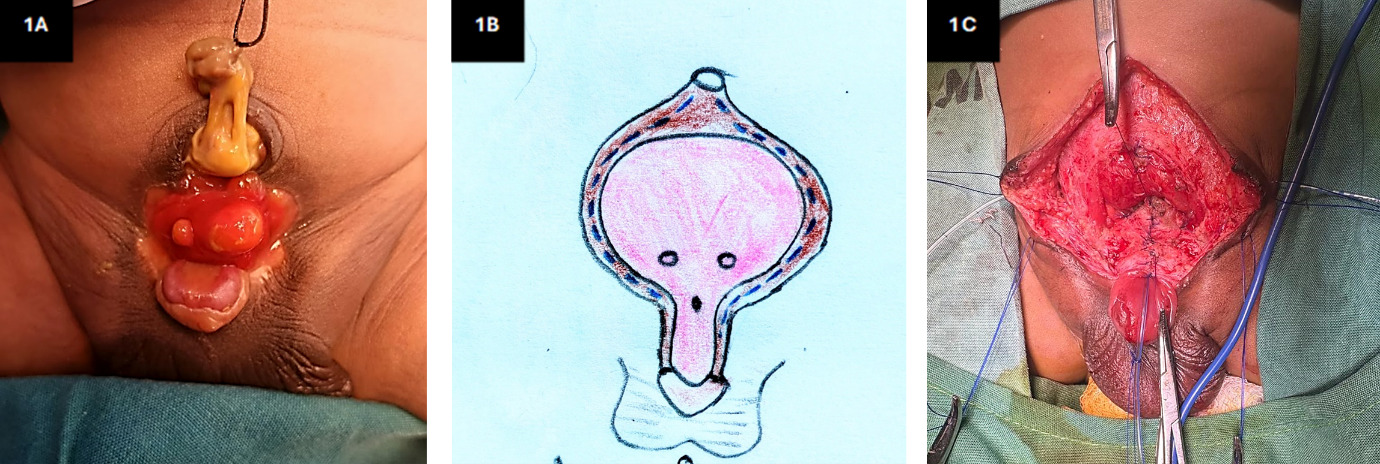
The incisions are deepened with monopolar diathermy, and careful dissection is carried out at a plane between the bladder plate anteriorly, the peritoneum posteriorly, and the rectus muscles laterally. At the level of the bladder neck, the intersymphyseal bands are incised to facilitate optimal placement of the bladder deep within the pelvis. The two ureteric orifices are intubated with a 4Fr stent, which will later be exteriorized, as shown in Figure 1C. The bladder and proximal urethra are reconstructed over a 6Fr urethral stent and an 8Fr suprapubic stent in 2 layers, an inner layer using a continuous absorbable monofilament 6/0 suture and an outer layer using a continuous or interrupted absorbable monofilament 5/0 suture. Bilateral herniotomy is routinely performed at this stage.
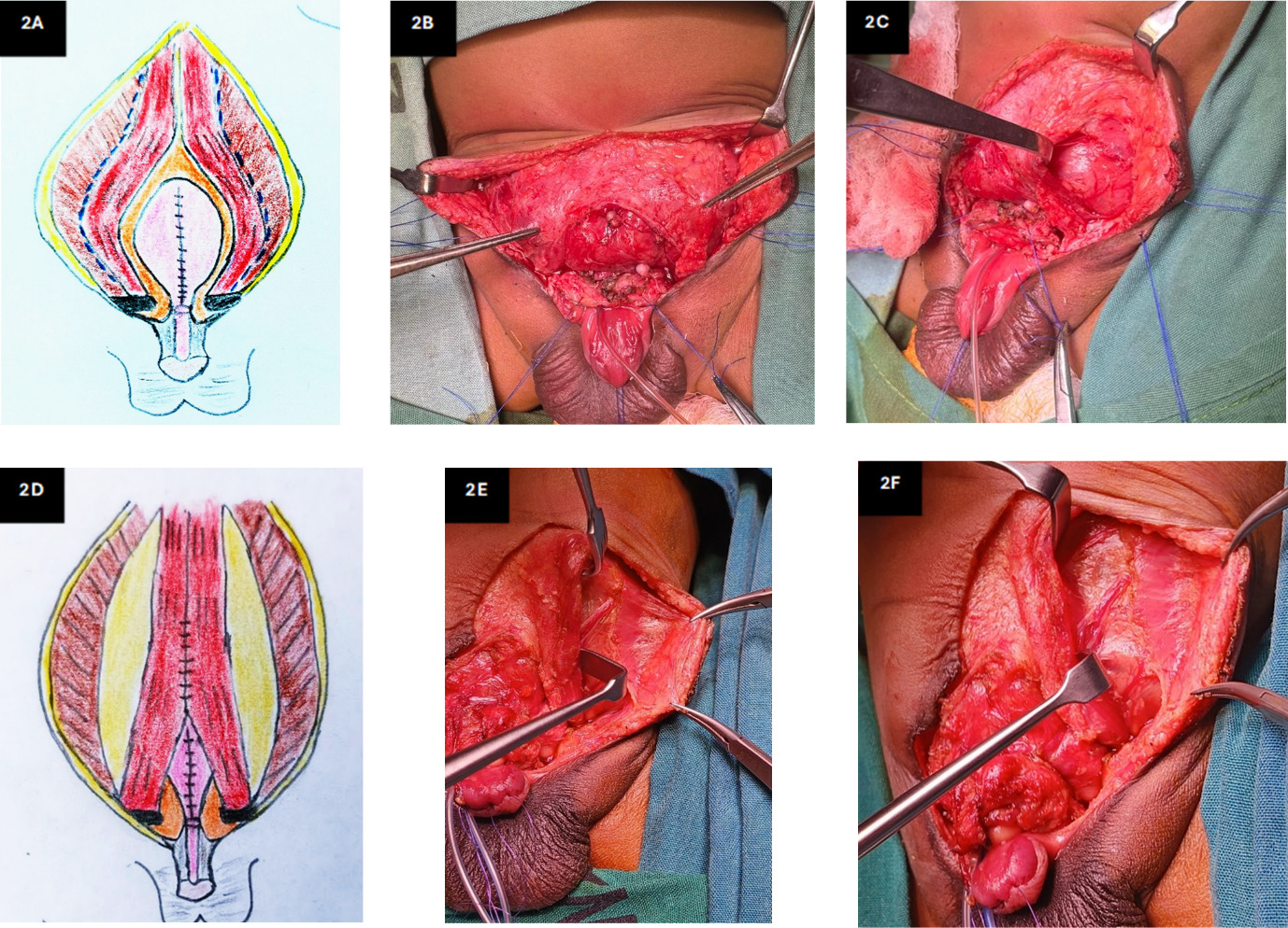
To achieve sufficient exposure to the rectus abdominis muscle, the incision is extended cranially in a longitudinal midline fashion to expose the superior confluence of the divergent rectus abdominis muscles. Mobilization of the rectus abdominis muscle is achieved by longitudinal dissection along the linear semilunaris, after which both anterior recti are mobilized medially while the external oblique aponeurosis and posterior rectus sheath rest laterally, as shown in Figure 2. Attention is taken to preserve the blood supply of the muscle during dissection and medial mobilization, as shown in Figure 2E.
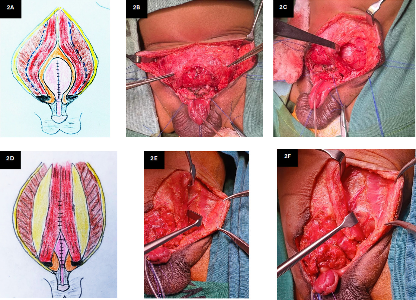
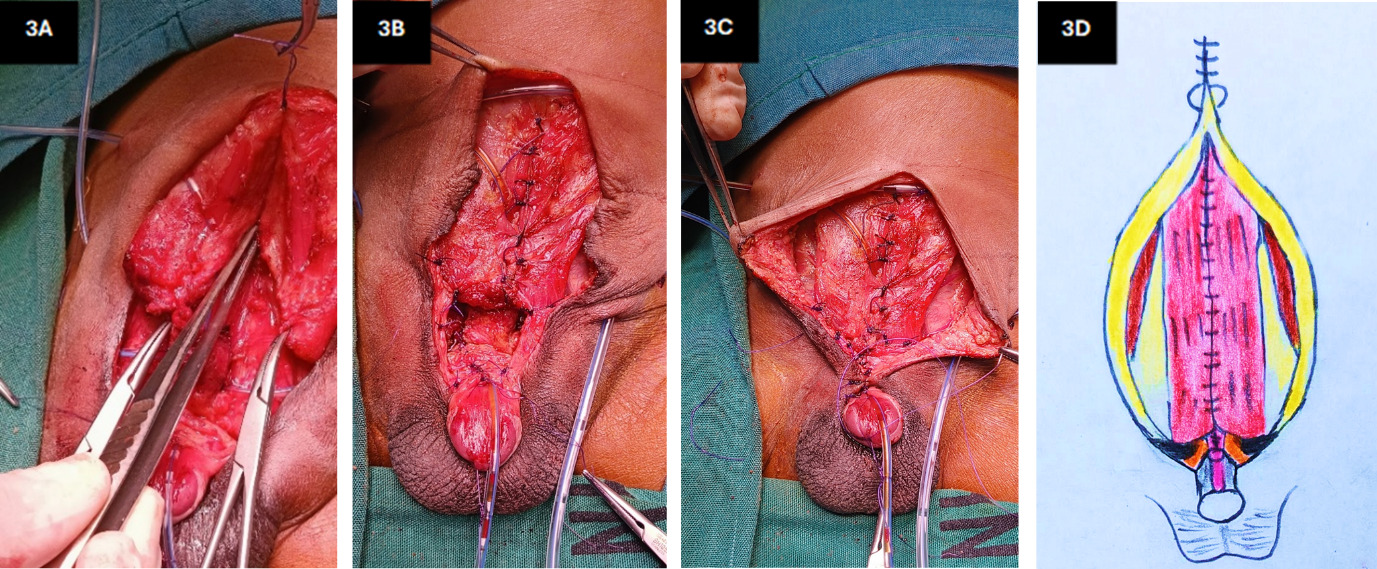
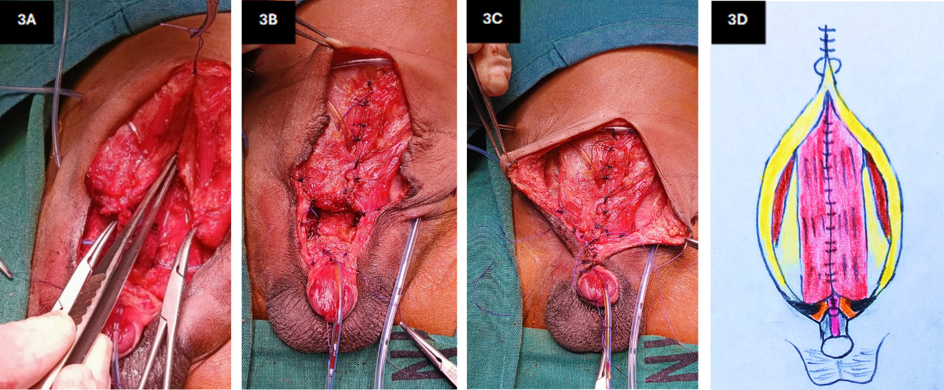
The two rectus abdominis muscles are then approximated over the bladder using interrupted absorbable monofilament or braided 3/0 or 2/0 suture as shown in Figure 3. The lower edge of the rectus remains anchored on the pubic bone while the distal medial rectus is approximated, facilitating abdominal wall closure.
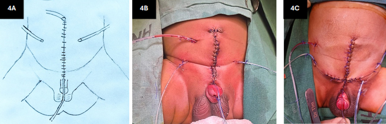
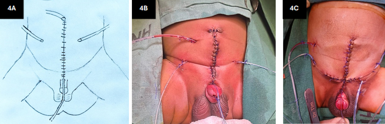
The two ureteral stents, a supra pubic stent and an optional subcutaneous drain, are exteriorized and secured to the skin, while the urethral stent is secured to the penile shaft as shown in Figure 4. The skin is closed with absorbable interrupted sutures (5/0 or 4/0), and a dressing is applied without pelvic immobilization.
Postoperatively, all the patients are nursed in the Surgical Neonatal Care Unit. Wound care is achieved by cleaning and dressing on an alternate-day basis. All patients are given antibiotics for 10 days and breastfed on demand. The drains and stents are removed as follows: drain on day 5; urethral stent on day 7; ureteral and suprapubic stents on day 21. The post-discharge follow-up schedule is 2-weekly until complete wound healing, then every 3 months for the first year, until epispadias repair, after which they are followed up 6-monthly until assessment for continence intervention is indicated.
Outcome
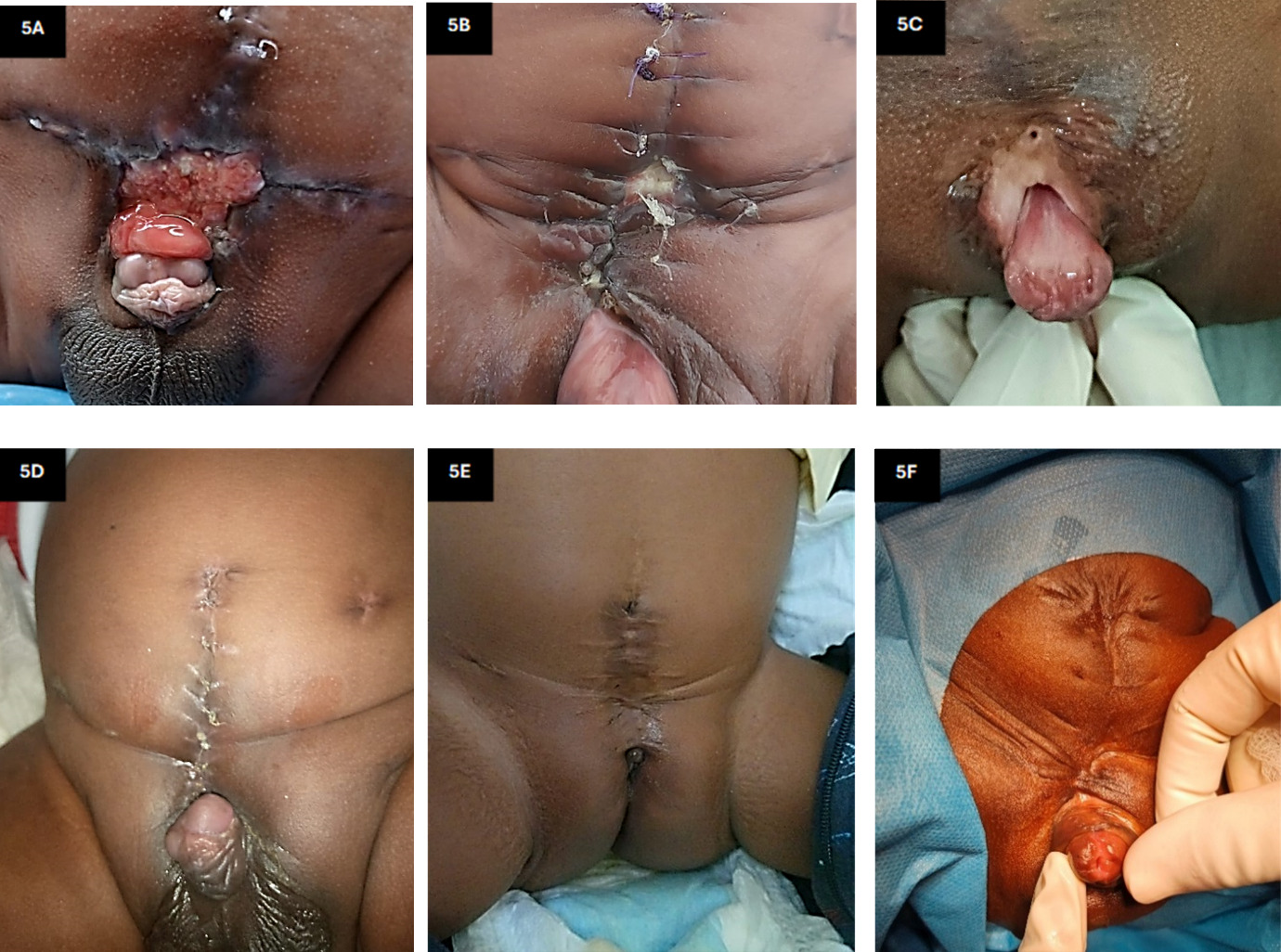
The mean length of inpatient hospital stay after surgery was 25 days. Complications occurred in 37.5% of patients: one had complete bladder and abdominal wall dehiscence (Figure 5A), one had a partial anterior abdominal wall dehiscence that healed by secondary intention (Figure 5B), and one developed a suprapubic fistula that resolved with conservative management (12.5%) (Figure 5C). The remaining 5 patients (62.5%) recovered without incident.
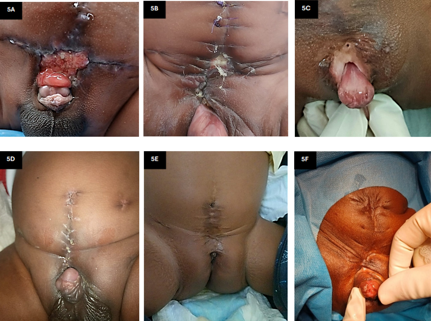
After discharge from the hospital, patients were followed up in the outpatient clinic according to the protocol. The patient with bladder dehiscence will be considered for redo surgery. The other 7 patients showed no abdominal wall herniations or defects (Figure 5D). The female patient (Figure 5E) also had a favourable outcome and is currently on serial bladder capacity measurements; she will be assessed for continence at a later stage. Five of the male patients have undergone epispadias repair, and one is shown in Figure 5F.
Discussion
Surgical management of bladder exstrophy is aimed at achieving a successful closure. Initially, repair was favoured within 72 hours to allow for pubic bone approximation without the need for osteotomy.2 However, current literature supports delayed closure with osteotomy, which ensures a tension-free approximation of the abdominal wall while allowing the bladder to sit deep within the pelvis.5 Conversely, some authors advocate for repair without osteotomy, due to osteotomy complication rates of 10%–30%, including pin-site infection, increased operative time (mean 45–90 additional minutes), greater intraoperative blood loss, and mandatory post-operative pelvic immobilisation for 4–6 weeks, which can lead to the development of pressure sores.4 Additionally, the recurrence of pubic diastasis after osteotomy has also been documented.3
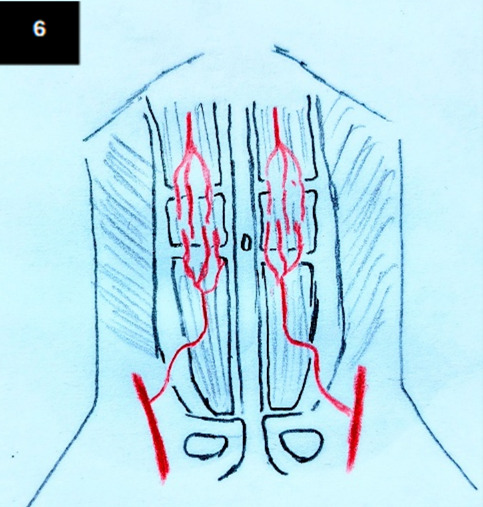
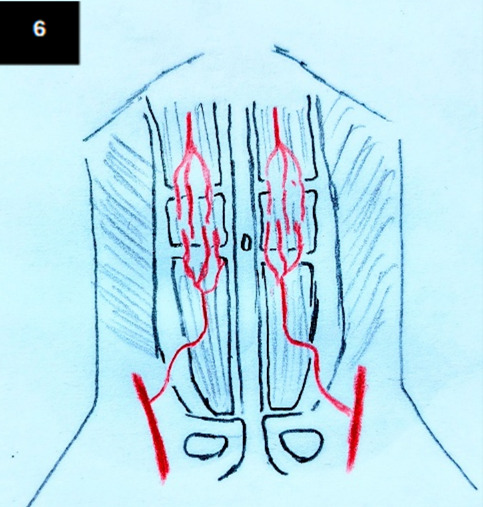
Rectus flap mobilization allows for closure of the bladder plate without the need for osteotomy. The principle of this repair is based on the attachments of the rectus abdominis muscle and its blood supply. The inferior attachment of the rectus abdominis muscle is to the pubic symphysis and crest, while laterally, the linear semilunaris defines a plane that separates the lateral border of the rectus abdominis from the medial border of the external oblique aponeurosis.6 In bladder exstrophy, there is pubic diastasis, and the rectus abdominis muscle is therefore divergent inferiorly as it approaches its attachments to the two pubic crests.7 The blood supply to the anterior abdominal wall is from the superior and inferior epigastric vessels, as shown in Figure 6. The inferior epigastric artery courses superiorly from the femoral artery and then courses medially to enter the rectus abdominis and anastomoses with the superior epigastric vessels around the umbilical region.
The understanding of this surgical anatomy is what enables the rectus muscle to be raised as a flap by separating the anterior rectus sheath and muscle from the posterior rectus sheath and its attachment to the external oblique aponeurosis at the level of the linea semilunaris. The two recti are then mobilized medially and approximated with minimal tension. In doing so, coverage of the anterior abdominal defect and the repaired bladder underneath is achieved. Notably, our technique is a slight modification from the previously described approaches by Hosseini et al, and Macedo et al in 2022.8,9 Hosseini et al describe mobilization of the rectus flap by dividing the inferior rectus attachments with a small pubic bone segment using diathermy, followed by upper fascial relaxing incisions on both rectus muscles, while Macedo et al describe a full anterior component separation technique that preserves the distal anchor and nerve supply during Kelly repair.8,9
The complications arising from primary repair of bladder exstrophy in MSRE are abdominal wall dehiscence, bladder dehiscence, bladder prolapse, and vesico-cutaneous fistula.10 Inability to achieve a tension-free repair is a major risk factor associated with the previously described complications. While an osteotomy is routinely offered in most repairs to mitigate tension, this approach of utilizing the rectus abdominis offers a reasonable alternative with an acceptable complication rate. Hosseini et al reported successful bladder closure in 13 patients, with only minor wound dehiscence in 3, without the need for osteotomy or pubic bone approximation.8 Similarly, Macedo et al in 2022 reported effective abdominal wall closure in 10 patients with no dehiscence or postoperative abdominal wall herniation; only 2 patients (20%) developed fistulae, both of which closed spontaneously.9 In our series, the dehiscence rate was slightly higher at 25%, which could be attributed to patient age at repair, limitations in wound care infrastructure, and the learning curve associated with early adoption of a modified technique. Although we observed no cases of denervation, rectus atrophy, distal muscle retraction, Spigelian hernia, or impaired continence in this case series, longer follow-up with formal electromyographic and imaging assessments is needed to better evaluate these potential complications.
Our approach to anterior abdominal wall closure in patients with bladder extrophy using a rectus abdominis flap seems more practical in our setting. Compared to pelvic osteotomies, it is not only less expensive but also requires a shorter hospital stay, is more suited to our surgical and nursing competencies, and reduces the social and economic disruption associated with poor follow-up. Because our case series is small, we are unable to assess the reliability of our findings or confirm the validity of this modified technique. Nevertheless, we suggest that future prospective, outcome-focused studies be conducted to further evaluate this approach.
Conclusion
This case series shows that our modified rectus mobilization approach for bladder exstrophy repair was successful in 7 of 8 cases. The main complications encountered were dehiscence in a single case and a fistula that closed spontaneously. The approach is practical and reproducible and can be used in settings where osteotomy is not feasible. This series describes our experience and short-term results; as such, this technique should be regarded as an institution-specific adaptation rather than a recommended alternative to established approaches. Our planned prospective follow-up, which incorporates bladder capacity measurements, continence assessment, abdominal wall imaging, and neuromuscular evaluation at defined intervals, is required to further validate the study.
Ethical Approval
Publication approval was granted by the KNH-UON Ethics Review Committee (Ref: KNH-ERC/01/PUB/6).
Informed Consent
All patients gave well informed consent for publication.
Data Availability
The data supporting the findings of this case series are included within the article. Additional de-identified data are available from the corresponding author upon reasonable request.
Conflict of Interest
None
Funding
None