Introduction
Peritonitis is inflammation of the peritoneum, which lines the abdominal cavity and its contained organs.1 Peritonitis can be classified into primary, secondary, or tertiary peritonitis. Secondary peritonitis, which is the most common, follows a pathologic process in an intra-abdominal viscus.2
Cytokines are small, non-structural proteins with molecular weights ranging from 8–40,0000d, which are synthesized by nucleated cells. They are primarily involved in host responses to disease and infection.3 Pro-inflammatory cytokines include interleukin (IL)-1, IL-8, IL-12, tumor necrosis factor (TNF), interferon (IFN) –γ, and granulocyte macrophage Colony stimulating factor. TNF-α is rapidly deployed in response to infection and is a potent mediator of the ensuing inflammatory response. It has a half-life of 18.2 minutes.4 IL-1 has 11 members, but IL-1β is the most studied member.3 It has a range of pro-inflammatory effects that are largely similar to TNF. It has a half-life of <6 minutes.5 These effects of either cytokine are greatly intensified by the presence of the other cytokine.6
In order to understand the time-course of inflammatory cytokine secretion, van Kralingen et al noted that most of the inflammatory response occurred within the initial 48-hour period, as most cytokines peaked by 48 hours. Levels of inflammatory cytokines returned to basal levels after 96 hours.7 Several prognostic indices are used to assess the severity of peritonitis. The Mannheim Peritonitis Index (MPI) is a widely used specific peritonitis score. It evaluates pre- and intra-operative clinical factors. These factors are organ failure, diffuse peritonitis, age older than 50 years, female gender, malignancy, non-colonic sepsis origin, pre-operative duration of peritonitis, and characteristics of the exudate (fecal, cloudy/purulent or clear).8 MPI > 25 is associated with poorer prognosis and increased mortality.9,10
This study assayed pro-inflammatory cytokines (TNF-α and IL-1β) in patients with secondary peritonitis with the aim of early risk stratification, prognostication and post-operative monitoring. The levels of TNF-α and IL-1β in apparently healthy individuals were also assayed.
Methods
This was a prospective longitudinal comparative study. Thirty-five consecutive consenting patients with secondary peritonitis presenting at the Emergency Department of the University College Hospital, Ibadan, Nigeria were prospectively enrolled.
The University College Hospital, Ibadan is Nigeria’s premier teaching hospital with over 1000 bed spaces. The location of the hospital is strategic as it serves Ibadan metropolis with referrals from the region. The hospital provides training, research, and health care services to the people of Nigeria. It has the full complement of medical and surgical specialties (and sub-specialties) with a modern, well-equipped emergency department and a functional intensive care unit.
Thirty-five patients without secondary peritonitis matched with the cases in sex, age (±5 years), and BMI (±3 kg/m2) served as controls. The study size was arrived at using the appropriate sample size formula while correcting for attrition. The period of recruitment was between January and December 2019, with data collection and follow-up lasting one year.
Blood samples were taken to assay for TNF-α and IL-1β at presentation once the diagnosis of secondary peritonitis was made and before resuscitation. Standard surgical procedure was performed for all participants after adequate resuscitation. 48 hours after surgery, blood samples were taken again for the estimation of these cytokines, representing post-resuscitation/intervention sample, while another sample was taken 96 hours after surgical intervention. Blood samples were also taken from the controls at presentation, 48 hours, and 96 hours after surgery. These samples were collected in 5 mL sterile tubes with lithium heparin anticoagulant and were centrifuged within 20 minutes at 1400g for 15 minutes. The supernatant plasma was separated and frozen at −40 oC until analysis. TNF-α and IL-1β were assayed using sandwich ELISA immunoassays (My BioSource, Inc., San Diego, CA, USA) on a Stat Fax 4000 ELISA reader (Awareness Technology Inc., Ramsey, USA).
These mediators were assayed by 1 laboratory scientist. All analysis was done in one analytical run. Quality control samples were included to determine analytical sensitivity, inter-assay variation, as well as intra-assay variation. The laboratory scientist was not aware of the patients’ characteristics to minimise assessment bias in the study. However, a degree of selection bias from the hospital based consecutive sampling of the cases was expected and unavoidable.
Data were collected and entered into a pro forma which was used for analysis. There were no missing data. Statistical analysis was carried out using the Statistical Package for the Social Sciences version 21. Pearson chi-square test was used to assess for the difference in categorical variables between the cases and the controls. Paired-samples T test and Mann-Whitney U test were used to assess the difference in parametric and non-parametric continuous variables between cases and the controls. The P value was considered to be significant if <0.05. Receiver operating characteristic curves showing the accuracy of TNF-α and IL-1β in predicting the severity of peritonitis were drawn. Repeated measure ANOVA curve for trends in observations of the participants was included. Multivariable binary logistic regression was used to identify independent predictors of post-operative mortality.
Sample size calculation
The minimum sample size was determined by the following formula, yielding a total of 70, including a 10% attrition rate.
Sample size:
Where,
n is the minimum sample size,
Zα is the critical value at 95% confidence interval = 1.96,
Z1-β is the power at 20% = 0.84,
SD is the standard deviation of TNF-α in healthy controls = 19 pg/mL,11
µ1 = mean value of TNF-α in healthy controls = 30 pg/mL,
µ2 = mean value of TNF-α in critically ill patients = 20 pg/mL.
Inclusion criteria
The study comprised patients aged 16 years and above with secondary peritonitis who had surgical intervention. Patients without secondary peritonitis matched with the cases in sex, age (±5 years), and BMI served as controls. These controls were patients who required laparotomy for conditions not associated with dysregulation of cytokines, such as trauma, inflammatory conditions/sepsis, and cancer.12 Such surgeries include laparotomy for non-septic and non-malignant causes, such as truncal vagotomy and pyloroplasty, repair of incisional hernia, and closure of colostomy.
Exclusion criteria
Individuals with factors that may be associated with falsely elevated cytokine levels, such as smokers,13 patients with Schizophrenia,14 patients on ACE inhibitors,15 and patients with depressed left ventricular ejection fraction.16
Ethical approval
Ethical approval was obtained from the Institute for advanced medical research and training (IAMRAT), College of Medicine, University of Ibadan IU/UCH ethics committee (UI/EC/17/0574). Utmost confidentiality was observed throughout the conduct of this research. This study did not adversely interfere with the duration or outcome of surgery, nor the length of hospital stay. Also, participation in this study was at no added cost to the patients. Informed consent for the study was obtained from the participants.
Results
One hundred and nine of the 7383 patients who were admitted to the emergency department during the year of the study period had secondary peritonitis. Secondary peritonitis accounted for 1.5% of all adult emergencies at this hospital. Thirty-five of these patients who consented to be included in the study were recruited as cases. Thirty-five other patients were recruited as controls. These controls were matched with the cases in sex, age (±5 years), and BMI (±3 kg/m2). Overall, 70 people were recruited.
Twenty-five (71.4%) of the cases were male, while 10 (28.6%) were female, giving a male-to-female ratio of 2.5:1. The mean age of the cases enrolled was 34.1, ranging from 16–62 years, while that of the controls was 37.9 years, with a range of 17–65 years. The modal age group for the participants of this study was 20–29 years. The mean BMI for the cases was 29.3 ± 3.42 kg/m2, while that of the controls was 32.7 ± 5.61 kg/m2. Most of the cases (57.1%) were of American Society of Anaesthesiologists (ASA) Class 3, while the controls were mostly (97.1%) either Class 1 or 2. Eleven cases (31.4%) required pre-operative transfusion, while none of the controls were transfused pre-operatively. Other characteristics are shown in Table 1.
The most common etiologies of secondary peritonitis in this series were complicated appendicitis 11 (31.4%), perforated PUD 10 (28.6%), and perforated typhoid ileitis 6 (17.1%). Other causes were strangulated adhesive intestinal obstruction 3 (8.6%), iatrogenic bowel injuries 3 (8.6%), and penetrating abdominal injury (5.7%). The procedures done for the cases in descending order were appendicectomy 11 (31.4%), closure of small bowel perforation 10 (28.6%), closure of gastric perforation 10 (28.6%), small bowel resection and anastomosis 3 (8.6%), and ileostomy 1 (2.9%), while the procedures done for the controls are closure of colostomy 17 (48.6%), repair of incisional hernia 14 (40%), repair of epigastric hernia 2 (5.7%), and truncal vagotomy and pyloroplasty 2 (5.7%). A vast portion (85.7%) of the cases had intra-operative findings of purulent peritoneal fluid, while 14.3% had fecal peritoneal fluid. The mean volume of peritoneal fluid was 1050 mL.
Four cases required blood transfusion post-operatively, while none of the controls did. Similarly, the cases that required intensive care, ventilator support, and inotropic support were 3, 2, and 2, respectively. None of the controls needed these. Two cases had tertiary peritonitis necessitating a relaparotomy. Secondary wound closure (2) for burst abdomen and left chest thoracostomy tube drainage (1) for sympathetic pleural effusion were additional procedures done, respectively. None of the controls had an additional procedure. The mean hospital stay for the cases was 12.9 (±8.0) days, while it was 6.4 days (±3.3) for the controls. The only mortality recorded during this study occurred on the 17th post-operative day for a case.
Repeated measure ANOVA curve for trends in temperature, pulse rate, and MAP showed a tendency to normal values over the period of hospitalization for the cases. This normalization is first noticeable about 48 hours after surgery. The controls invariably had normal values throughout admission. The incidence of post-operative complications such as surgical site infection, enterocutaneous fistula, and wound dehiscence was more common in cases compared with controls.
The plasma levels of TNF-α in cases pre-operatively, 48 hours post-operatively, and 96 hours post-operatively were 192.2, 213.3 and 217.1 pg/ml respectively while the levels for controls were 12.92, 13 and 12.9 pg/ml respectively. The plasma levels of IL-1β in cases pre-operatively, 48 hours post-operatively, and 96 hours post-operatively were 9, 8.7 and 8.9 pg/ml respectively while the levels for controls were 7.7, 7.1 and 6.5 pg/ml respectively. TNF-α showed a 15-fold elevation between cases and controls at each measured point, while IL-1β showed a 1.2-fold elevation. The plasma levels of pro-inflammatory cytokines (TNF-α and IL-1β) are higher in patients with secondary peritonitis at each time limits (P = 0.28). This difference was not statistically significant.
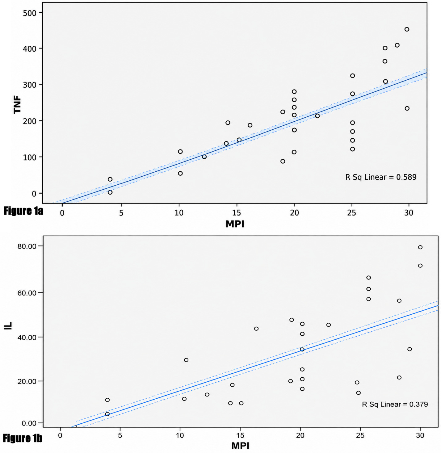
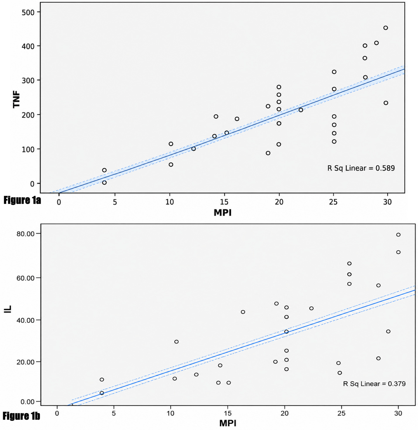
The relationship between the values of cytokines and the Mannheim Peritonitis Index is linear and positive, meaning higher MPI values were associated with higher values of the cytokines assessed. This relationship was represented in scatter diagrams in Figure 1A and B.
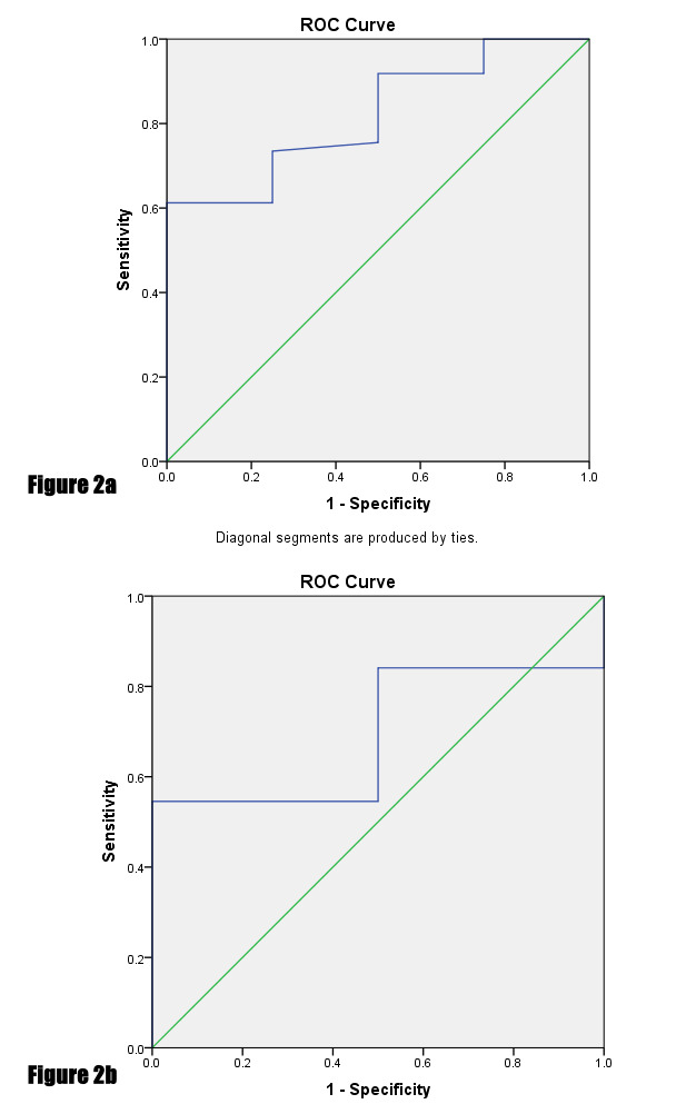
A cut-off value of 21.75 pg/mL for TNF-α had a sensitivity of 95% and a specificity of 90% (Figure 2A), while a value of 6.47 pg/ml for IL-1β had a sensitivity of 73% and a specificity of 67% (Figure 2B). The values of 21.75 and 6.47 have the highest combined sensitivity and specificity for TNF-α and IL-1β, respectively. The binary outcome for receiver operating characteristic (ROC) analysis was MPI > 25, and this positively correlated with the presence of post-operative complications such as surgical site infection, enterocutaneous fistula, wound dehiscence, ICU admission, and mortality.
_roc_curve_showing_the_accuracy_of_tnf-_in_predicting_severity_of_peritonitis_(auc__ar.png)
Multivariable binary logistic regression was used to identify independent predictors of post-operative mortality. Mortality was the dependent variable, while ASA score > 3, MPI score > 25, and post-operative enterocutaneous fistula were identified as significant covariates. Cases with post-operative complications had higher mean levels of TNF-α and IL-1β compared with those without complications (P = 0.34). These post-operative complications included surgical site infection, enterocutaneous fistula, wound dehiscence and mortality. The difference in levels was not statistically significant. The comparison of mean cytokine levels of cases with significant peri-operative events is summarized in Table 2.
Of the 35 cases, 26 had a positive peritoneal fluid culture. The common isolates are Escherichia coli (38%), Klebsiella spp (23%), Enterococcus faecalis (19%), Enterobacter cloacae (12%), Pseudomonas spp (8%), and Bacteroides spp (23%). A single pathogen was isolated in 18 (69%) cases, whereas 8 (31%) cultures identified multiple pathogens.
Mean values of TNF-α and IL-1β were higher in those with a positive peritoneal fluid culture. The values were 305.5 and 31.4 pg/mL, respectively, in individuals with a positive culture, while those whose peritoneal culture yielded no growth had values of 176.9 (P = 0.002) and 30.4 pg/ml (P = 0.613), respectively. The values of these cytokines were comparable for all organisms irrespective of the type; however, values were colossally higher in multiple isolates. The values of TNF-α and IL-1β for those with multiple isolates were 364.0 and 53.2 pg/mL, respectively, compared with 255.7 (P = 0.001) and 26.3 (P = 0.002) pg/mL in those with a single isolate.
Discussion
The plasma levels of TNF-α and IL-1β of the cases were consistently higher than those of the controls at all points (at admission, 48- and 96-hours following surgery). The range of TNF-α and IL-1β that was obtained from this study for the controls is 5.85–60.70 pg/mL and 5.98–19.63 pg/mL, respectively. Reference ranges of cytokines, however, vary considerably depending on age, sex, and disease state.17 Pro-inflammatory cytokines have been established to be elevated in secondary peritonitis.18–20
Secondary peritonitis is a prevailing surgical emergency, especially in developing countries.21 It accounted for 1.5% of all adult emergencies at this hospital during the study period. Worldwide, secondary peritonitis represents about 1% of emergent hospital admissions.1 The slightly higher percentage seen in this hospital may be because of its tertiary healthcare status, which subserves the state and its environs. All cases presented as emergencies, while most of the controls (97.1%) were electives.
The ASA classification is a simple and reliable predictor of complications and mortality following surgery.22 A greater number of the cases (57.1%) were of ASA Class 3, while almost all controls (97.1%) were either Class 1 or 2. Most subjects with secondary peritonitis in other studies23,24 also had an ASA Class of 3. Similarly, studies25,26 that have evaluated the outcome of patients undergoing elective surgery in this hospital have most of the cases to be either ASA 1 or 2.
Repeated measure ANOVA curve for trends in temperature, pulse rate, and MAP showed a tendency to normal values over the period of hospitalization for the cases. This normalization is first noticeable about 48 hours after surgery. The controls invariably had normal values throughout admission.
The prevailing etiologies of secondary peritonitis in this study are complicated appendicitis, perforated PUD, and perforated typhoid ileitis. These are the commonest causes of secondary peritonitis in adults,21,23 especially in the developing world. The etiology of non-traumatic secondary peritonitis in developing countries, being proximal bowel perforation, is consistent with reports.23,27
The mean volume of fluid administered, the units of blood transfused, and blood loss are established surrogates of the complexity of surgery28 while they also mirror the magnitude of haemodynamic instability and third space loss.23 These were higher in surgeries for cases than in the controls. Surgical management of secondary peritonitis is associated with more meticulous use of intravenous fluids and blood products.29
A review of the temporal variation in values of TNF-α over 96 hours showed a slight increase for cases but remained relatively constant for controls. The opposite was the case for IL-1β, in that the elevated values were sustained for the cases over the period of study (96 hours). The control population demonstrated a decline over the same period. This suggests that surgical trauma alone does not contribute to a cytokine storm. A study evaluating the changes in serum cytokines in response to musculoskeletal surgical trauma30 did not show a direct correlation between TNF-α and IL-1β with the degree of injury. The ROC curve for TNF-α showed better characteristics with a sensitivity of 95% and a specificity of 90% at a cut-off value of 21.75. A sensitivity of 73% and a specificity of 67% for IL-1β were obtained from this study. TNF-α also showed higher sensitivity and specificity than IL-1β in primary peritonitis31 and sepsis.32
Complications such as surgical site infection, enterocutaneous fistula, and wound dehiscence were higher in the cases. The overall rate for surgical site infection in this study was 48.6%. Surgeries for peritonitis and emergency surgeries have a high rate of surgical site infection.33 The mortality rate for the cases was 2.9%, and significant risk factors identified using multivariable binary logistic regression analysis include high ASA (>3) & MPI (>25) scores and post-operative enterocutaneous fistula. The values of TNF-α and IL-1β were significantly higher in the case of mortality as opposed to survivors.
The levels of TNF-α and IL-1β were elevated in individuals with post-operative complications compared with those without. These cytokines have been established to be higher in post-operative inflammatory or infective complications. Lower levels of cytokines are significantly associated with a favorable outcome.22 In this study, higher levels of TNF-α and IL-1β were measured in patients who had relaparotomy. TNF-α and IL-1β levels were also higher in non-survivors, and this is congruent with findings seen in other studies. Peritonitis precipitates an acute systemic and peritoneal innate immune response with a concurrent release of pro-inflammatory cytokines. Higher levels of cytokines are observed in the plasma of patients with the most severe conditions such as those in shock and non-survivors.18
The role of cytokines in wound healing suggests an elevation in levels following secondary wound closure. The repair process after injury is initiated after the release of various growth factors, cytokines, and low-molecular-weight compounds from the serum of damaged blood vessels and platelets. Expression of TNF-α and IL-1β is categorically upregulated during the inflammatory phase of healing.34
Anemia is associated with higher cytokine levels. TNF-α and IL-1β are associated with anemia by truncating red cell survival, blunting erythropoietin response to anemia, and anomalous mobilization of reticuloendothelial stores. An account of higher cytokine levels in those requiring blood transfusion is also reported.35
The mean return of bowel function was higher in the cases with higher cytokine levels. Although an integrated understanding of the pathophysiology of post-operative ileus is elusive, some mechanisms have been described. Factors that can specifically contribute to post-operative ileus following secondary peritonitis include the intraperitoneal nature of surgery, exaggerated inflammatory response, greater degree of bowel manipulation, and extended duration of operation. Post-operative ileus contributes significantly to morbidity, prolonged hospitalization, and increased health care expenditure. Pro-inflammatory cytokines such as IL-1β, IL-6, TNF-α, and CRP in serum and abdominal exudates play a pivotal role in the development of ileus following abdominal surgery.36
In this cohort, the proportion of positive peritoneal fluid cultures was 74%. Higher levels of cytokines were associated with polymicrobial culture; however, these values were comparable for all organisms irrespective of the genus. Riché et al18 noted higher levels of TNF-α in the presence of peritoneal anaerobes or Enterococcus species.
Limitations
Despite the expanding body of research, some limitations must be acknowledged. There are few studies on cytokines in peritonitis in human subjects especially in Sub-Saharan Africa. Assay of pro-inflammatory cytokines in this study did not approximate values of the apparently healthy controls, even 96 hours after surgery, despite the half-lives for TNF-α and IL-1 being 18.2 minutes4 and <6 minutes,5 respectively. This provokes the question of when levels of these cytokines return to normal or pre-morbid levels. The kinetics and threshold values of these cytokines are knowledge gaps that remain. A follow-up study would involve assay of all pro-inflammatory cytokines (and perhaps anti-inflammatory cytokines as well) at a much shorter frequency, including prior to discharge and at least once during the follow-up period at the outpatient clinic, while including survival analysis preferably in a large-scale and multi-center investigation.
Conclusion
The plasma levels of pro-inflammatory cytokines (TNF-α and IL-1β) are higher in patients with secondary peritonitis at admission, 48 hours, and 96 hours following surgery as compared with apparently healthy controls. The mean values of TNF-α and IL-1β in cases with post-operative complications are higher than in those without.
Pro-inflammatory cytokines could potentially aid early diagnosis, risk stratification, prognostication, and monitoring of therapeutic responses of patients with secondary peritonitis. However, the current data from this study are insufficient to support routine clinical implementation of TNF-α or IL-1β testing in patients.
Ethical approval
Ethical approval was obtained from the Institute for advanced medical research and training (IAMRAT), College of Medicine, University of Ibadan IU/UCH ethics committee (UI/EC/17/0574).
Informed Consent
Informed consent for the study was obtained from the participants
Data Availability
All data are available on reasonable request.
Conflict of Interest
None
Funding
None